Yes, many cases of reduced cervical lordosis can be improved non-surgically. The key is combining targeted cervical extension traction with a multimodal rehabilitation program, not just stretching or generic exercise. A systematic review of controlled trials found average radiographic lordosis increases of roughly 12–18° after extension traction protocols lasting 5–15 weeks, with traction groups maintaining pain and disability improvements at follow-up, while comparison groups without traction often regressed within a year.
That said, not every case responds equally. Rigid, degenerative deformities or cases with progressive neurological deficits may require surgical evaluation, as Cleveland Clinic guidance makes clear. The practical starting point for most people is a lateral cervical X-ray to establish baseline C2–C7 metrics, followed by a conversation with a clinician about whether an 8–12 week supervised traction and exercise program is appropriate.
- Get a lateral cervical radiograph to measure your C2–C7 Cobb angle and anterior head translation before starting any program.
- Discuss cervical extension traction options (Denneroll, 3-point bending traction, or CBP-style protocols) with a qualified clinician.
- Expect a supervised program of roughly 3 sessions per week for 8–12 weeks, combined with daily home orthotic use.
- Rule out red flags first: progressive weakness, gait disturbance, or bowel/bladder changes require medical evaluation before any corrective program.
Pro Tip: Bring a printed copy of your lateral cervical X-ray report to your first appointment. Clinicians who use radiographic endpoints can set a measurable goal in degrees, which gives you something concrete to track rather than just “feeling better.”
Key Takeaways
Cervical extension traction combined with multimodal rehabilitation is the most evidence-supported approach to structural cervical lordosis restoration, producing average radiographic gains of 12–18° over 8–15 weeks in controlled trials.
| Point | Details |
|---|---|
| Traction is the differentiating variable | Multimodal rehab without extension traction typically regresses within a year; traction groups maintain gains. |
| Expect 12–18° average radiographic gain | Controlled trials report this range over 5–15 weeks; single-case intensive protocols have shown larger gains. |
| Maintenance care is required | Partial regression occurs without ongoing home orthotic use and roughly twice-monthly maintenance visits. |
| Red flags need medical evaluation first | Progressive weakness, gait changes, or bowel/bladder dysfunction require clinical assessment before any corrective program. |
| Bestforwardheadposturefix | Provides sample 8–12 week program frameworks, clinician checklists, and exercise guides to support informed clinical care. |
Table of Contents
- What does the research actually show about restoring cervical lordosis?
- How clinicians measure cervical lordosis and track your progress
- What conservative methods can restore cervical lordosis?
- What does an evidence-backed treatment program actually look like?
- Who benefits from non-surgical restoration, and what are the red flags?
- A sample 8–12 week clinic and home program to discuss with your clinician
- What are the real limits of cervical lordosis restoration?
- Safety, contraindications, and questions to ask before you start
- An editorial perspective on what most people get wrong about cervical lordosis restoration
- What Bestforwardheadposturefix offers readers who want a structured starting point
- Sources
What does the research actually show about restoring cervical lordosis?
The strongest evidence comes from a systematic review of controlled trials examining cervical extension traction as part of rehabilitation. Across trials spanning 5–15 weeks and 15–60 treatment sessions, traction groups gained an average of 12–18° of lordosis on lateral radiographs. More telling: at one-year follow-up, traction groups maintained their pain and disability improvements, while comparison groups receiving multimodal rehab without specific extension traction had largely regressed.
Randomized trials using the Denneroll cervical traction orthotic add another layer of detail. Studies summarized by Oakley et al. showed that adding the Denneroll to a multimodal program produced superior radiographic correction on the absolute rotation angle (ARA) C2–C7 and reduced anterior head translation (AHT) compared with multimodal care alone. Some of those advantages held at one-year follow-up. The same trials also reported improvements in sensorimotor and autonomic measures, suggesting that structural correction may affect more than local pain.
CBP (Chiropractic BioPhysics) case reports document gains exceeding 20° in some patients using mirror-image exercises, cervical extension traction, and spinal manipulation together. A Pettibon protocol case report described a 144% increase in cervical lordosis and a 56% reduction in forward head posture after 30 sessions over 8 weeks, with symptomatic relief beginning around week 3. These are single-case examples, not generalizable trial data, but they illustrate what intensive multimodal protocols can produce in motivated patients.
The honest caveat: most trials combine multiple interventions, sample sizes are often modest, and protocols vary enough that direct comparison is difficult. Radiographic verification is essential because symptom improvement does not reliably track structural change.
How clinicians measure cervical lordosis and track your progress
Clinicians use lateral cervical radiographs as the primary measurement tool. The two most common metrics are the C2–C7 Cobb angle and the absolute rotation angle (ARA). The Cobb method draws lines along the inferior endplates of C2 and C7 and measures the angle between them; the ARA uses posterior tangent lines. A normal lordotic curve typically falls in the range of 20–40° by Cobb measurement, though published norms vary by method and population. A reduced or reversed (kyphotic) angle is the radiographic signature of hypolordosis.
Anterior head translation (AHT) measures how far the center of the head sits in front of the C7 plumb line. Elevated AHT is a common companion to loss of lordosis and is independently associated with increased mechanical load on the cervical spine. Trials using the Denneroll reported reductions in AHT alongside lordosis gains, which matters because forward head position and curve loss tend to reinforce each other.
Functional outcome measures like the Neck Disability Index (NDI) and a visual analog scale (VAS) for pain are used alongside imaging because structural and symptomatic improvement do not always move in lockstep. A patient can feel significantly better before radiographic change is detectable, or conversely, show radiographic improvement while still reporting disability.
Re-imaging is typically scheduled around the 8–12 week mark, after an initial treatment block. Clinicians generally avoid more frequent imaging to limit cumulative radiation exposure. Some practices use digital X-ray systems with lower dose protocols specifically for this reason.
Pro Tip: Ask your clinician which measurement method they use (Cobb vs. ARA) and request a printed copy of your baseline and follow-up measurements. Having the actual degree values lets you track progress objectively rather than relying on subjective impression alone.
What conservative methods can restore cervical lordosis?
Passive stretching and basic neck exercises alone rarely produce reliable structural correction. The literature consistently points to a multimodal approach as the standard for meaningful cervical curve restoration.
Cervical extension traction orthotics
The Denneroll and 3-point bending traction devices apply a sustained extension load to the cervical spine, targeting viscoelastic deformation of spinal ligaments and soft tissues over time. The Denneroll is a contoured foam orthotic placed under the neck while the patient lies supine; the head’s weight provides the corrective force, with duration and positioning adjusted progressively. Randomized trials show this approach produces superior radiographic and functional outcomes compared with multimodal rehab alone. CBP-style 3-point bending traction uses a similar principle with a more structured mechanical apparatus, often in a clinic setting.
Targeted exercise approaches
Mirror-image cervical extension exercises, chin tuck progressions, and thoracic extension drills address muscle imbalance and motor control. Cervical retraction exercises activate deep cervical flexors and reduce the forward head position that accompanies hypolordosis. These exercises are necessary for neuromuscular retraining but produce limited structural change on their own without the sustained mechanical load that traction provides.

Manual therapy and spinal manipulation
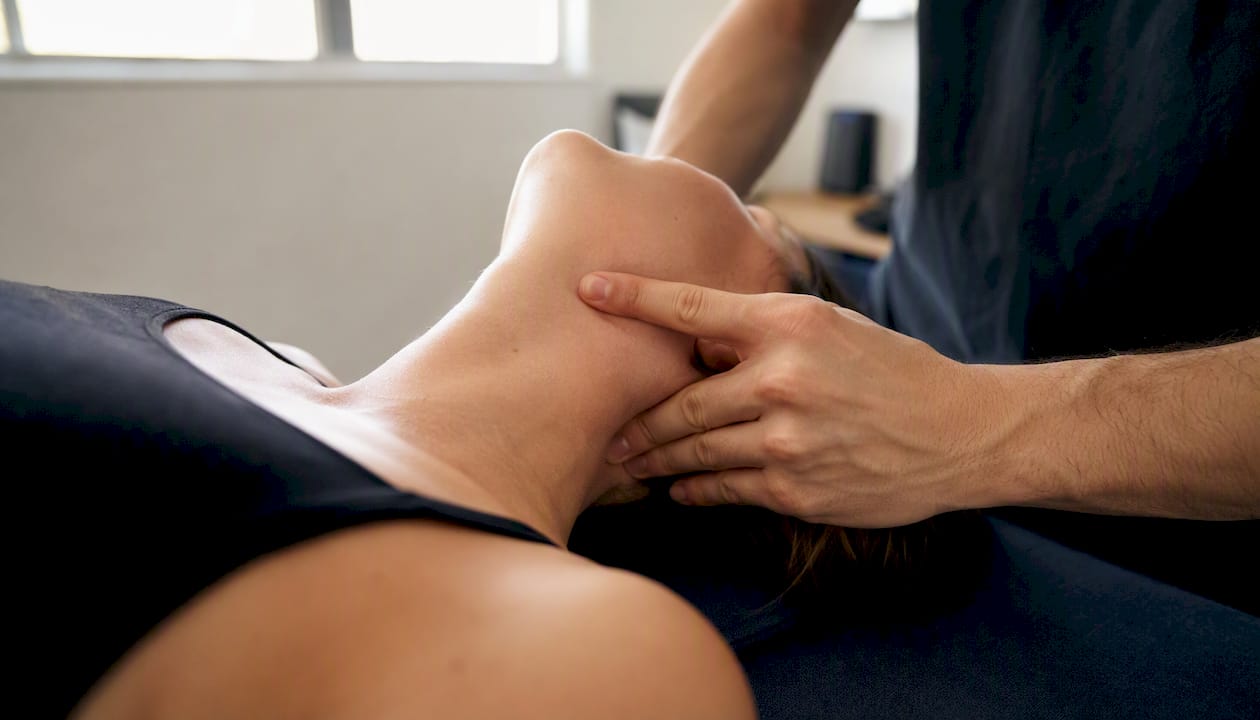
Spinal manipulation and manual therapy offer short-term pain relief and can reduce muscle guarding enough to make corrective loading more tolerable. Chiropractic care can also help restore range of motion that facilitates traction positioning. The evidence does not support manipulation alone as sufficient for sustained lordosis change, but as an adjunct it plays a clear facilitative role.
Home orthotics and progressive loading
Home use of the Denneroll or similar devices allows daily corrective loading between clinic visits, which is critical for consolidating viscoelastic changes. Clinical protocols in the literature escalate traction dosing progressively, starting with shorter durations and increasing as tolerance builds. Adherence to daily home sessions is one of the strongest determinants of outcomes in published trials.
| Method | Effectiveness (lordosis change) | Typical frequency/duration | Clinician supervision | Device cost | Evidence level |
|---|---|---|---|---|---|
| Cervical extension traction (Denneroll/CBP) | 12–18° average in systematic review | 3×/week clinic + daily home; 8–15 weeks | Required for setup; home use after training | Moderate (device purchase) | RCTs, systematic review |
| Mirror-image exercises + chin tucks | Modest alone; significant when combined | Daily; 8–12 weeks | Initial supervision recommended | Minimal | RCTs (as part of multimodal) |
| Manual therapy/SMT | Pain/mobility relief; not structural alone | 1–3×/week adjunct | Clinician required | Per-visit fee | RCTs (adjunct role) |
| Thoracic extension drills | Supports global alignment; indirect effect | Daily home | Self-directed after instruction | None | Clinical consensus |
| Home orthotic progressive loading | Consolidates clinic gains | Daily; ongoing | Periodic check-ins | Moderate (device) | Case series, RCTs |
Pro Tip: If your clinician prescribes home Denneroll use, set a phone timer for each session and log your duration daily. Adherence tracking in trials consistently separates patients who maintain gains from those who regress.
What does an evidence-backed treatment program actually look like?
Most published trials that produced meaningful lordosis gains used a frequency of roughly 3 clinic sessions per week for 8–12 weeks, totaling 30–40 sessions. Daily home orthotic use ran alongside clinic visits throughout. That schedule is demanding, and adherence is a major determinant of outcomes.
A typical week in the clinic phase looks like this: a traction session (Denneroll or 3-point bending, progressively dosed), manual therapy or spinal manipulation to reduce guarding and improve segmental mobility, and targeted exercise (mirror-image extension, chin tucks, thoracic extension). At home, patients use the orthotic daily for a prescribed duration that increases over the program, plus a short exercise set.
Radiographic change of 12–18° is the range reported in the systematic review across controlled trials. The Pettibon case report described a 144% increase in cervical lordosis after 30 sessions over 8 weeks, though that is a single-case result, not a typical expectation.
Symptom improvement often precedes structural change. Pain and NDI scores frequently improve within the first 3–4 weeks, which can mislead patients into thinking the structural work is done. It is not. Radiographic re-assessment at 8–12 weeks is the only reliable way to confirm whether the curve has actually changed.
After the initial program, maintenance care typically drops to roughly twice per month, with continued home orthotic use on a tapered schedule. CBP follow-up data shows that without some form of maintenance, initial corrections can partially regress over months to years.
Pro Tip: Schedule your 8–12 week re-imaging appointment before you start the program, not after. Patients who have a concrete re-assessment date on the calendar tend to maintain adherence better through the middle weeks when motivation dips.
Who benefits from non-surgical restoration, and what are the red flags?
The best candidates for non-surgical cervical lordosis restoration share a few characteristics: a flexible (non-rigid) deformity, mechanical neck pain without progressive neurological deficit, and the practical capacity to commit to a 3×/week clinic schedule plus daily home work. Baseline imaging confirming hypolordosis or mild kyphosis without severe multilevel degeneration is also a prerequisite for setting realistic goals.
Candidate profile:
- Flexible hypolordosis or mild cervical kyphosis confirmed on lateral X-ray
- Mechanical neck pain, headaches, or upper back tension without neurological signs
- No prior cervical fusion at the levels being treated
- Motivated and able to attend frequent clinic visits and maintain daily home protocols
- Cleared by a clinician after neurological examination and baseline imaging
Red flags that require medical or surgical evaluation before any corrective program:
- Progressive muscle weakness in the arms or hands
- New or worsening gait disturbance or balance problems
- Bowel or bladder dysfunction (urgency, incontinence, retention)
- Rapidly worsening neurological signs (numbness, tingling that is spreading)
- Signs of infection: fever, night sweats, unexplained weight loss with neck pain
- Severe or rigid multilevel ankylosis on imaging
- Advanced myelopathy signs on clinical exam
Conditions that substantially limit non-surgical success include rigid ankylosis from advanced degenerative fusion, severe myelopathy, and unstable fractures. For these, cervical fusion surgery may be the appropriate path to restore alignment and protect the spinal cord.
Questions to bring to your clinician: Can you perform a flexibility assessment to determine whether my deformity is rigid or mobile? What does my baseline imaging show about segmental mobility? Do I have any neurological findings that would change the plan?
Pro Tip: A simple in-office flexibility test (passive cervical extension range of motion) gives your clinician a quick read on whether the curve has any mobility to work with. If passive extension is severely restricted, that changes the prognosis for non-surgical correction significantly.
A sample 8–12 week clinic and home program to discuss with your clinician
This outline is a starting framework for a conversation with your provider, not a self-directed prescription. Traction dosing and exercise progression must be supervised and individualized.
Weeks 1–2: Baseline and introduction
- Clinic visit (3×/week): 10–15 minutes cervical extension traction at initial low load; 10 minutes manual therapy or soft-tissue work; 10 minutes supervised exercise.
- Home: Denneroll or prescribed orthotic for 10–15 minutes daily; chin tucks 2 sets of 10 reps, twice daily.
- Clinician establishes baseline NDI and pain VAS scores; reviews lateral cervical X-ray measurements.
Weeks 3–6: Progressive loading
- Clinic: Traction duration increases to 20–30 minutes per session; manual therapy continues as needed; add resisted chin tucks with a light band (2–3 sets of 12 reps) and thoracic extension over a foam roller (2 sets of 10 reps).
- Home: Orthotic time increases to 20–25 minutes daily; continue chin tuck sets; add mirror-image cervical extension exercise (2 sets of 10 reps, holding 3–5 seconds each).
- Progression rule: advance duration or load only if no new neurological symptoms appear and pain does not worsen beyond a transient 1–2 point VAS increase that resolves within 24 hours.
Weeks 7–12: Consolidation and re-assessment
- Clinic: Maintain traction at therapeutic dose; shift manual therapy to less frequent adjunct; introduce postural retraining for workstation and sleep positioning.
- Home: Maintain orthotic schedule; add posture correction habits for screen time and sleeping position.
- Re-imaging at week 8–12 to quantify radiographic change; re-score NDI and VAS.
Safety notes and stop criteria:
- Stop traction immediately if new numbness, tingling, or weakness develops in the arms or hands.
- Stop and seek medical review for sudden severe headache, visual changes, or dizziness that does not resolve within minutes of ending a session.
- Reduce traction duration (not just load) if symptoms flare beyond the 24-hour tolerance window.
- Clinician checklist: document traction dose at each session, track neurological screen at each visit, and confirm home orthotic compliance at each check-in.
Pro Tip: Mirror-image exercises work best when performed immediately before traction, not after. The pre-activation of extensors primes the musculature to hold the corrective position during the traction load, which may improve the neuromuscular component of the correction.
What are the real limits of cervical lordosis restoration?
Not every patient achieves full radiographic normalization, and the evidence base has genuine gaps worth understanding before you commit to an intensive program.
- Research limitations: Most trials combine multiple interventions simultaneously, making it difficult to isolate the contribution of any single component. Sample sizes in many studies are modest, and some of the larger reported corrections come from case reports rather than controlled trials.
- Partial correction is the norm: Trials report average gains of 12–18°, not full normalization to a textbook 30–40° curve. Patients with more severe baseline deformity or longer duration of loss tend to achieve less complete correction.
- Rigid deformities respond poorly: Advanced multilevel degenerative fusion limits the viscoelastic deformation that traction depends on. If segmental mobility is severely restricted at baseline, structural gains will be modest regardless of protocol intensity.
- Maintenance is not optional: CBP follow-up data consistently shows partial regression of initial corrections without structured maintenance. Twice-monthly maintenance visits and continued home orthotic use are commonly recommended after the initial correction phase.
- Symptom relief and structural correction are not the same thing: Feeling better is real and valuable, but it does not confirm that the curve has changed. Imaging is the only way to verify structural progress. Conversely, some patients show radiographic improvement without proportional symptom relief, which is why both imaging and functional scores matter.
Pro Tip: If your clinician does not plan to re-image at the end of your initial program, ask why. Radiographic follow-up is the only objective way to determine whether the structural goal was met and whether maintenance care is warranted.
Safety, contraindications, and questions to ask before you start
Cervical extension traction is safe for most patients when properly supervised, but specific contraindications make it inappropriate or dangerous for some.
Absolute contraindications to cervical traction or aggressive extension:
- Unstable cervical fractures or ligamentous instability (e.g., dens fracture, atlantoaxial instability)
- Acute inflammatory arthritis affecting the cervical spine (e.g., active rheumatoid involvement of C1–C2)
- Uncontrolled osteoporosis with high fracture risk
- Recent cervical fusion at the levels being tractioned
- Active infection, tumor, or vascular anomaly in the cervical region
Warning signs during treatment that require immediate stop and medical review:
- New or worsening numbness, tingling, or weakness in the arms, hands, or legs
- Bowel or bladder changes (urgency, incontinence, or retention) that develop during or after sessions
- Sudden severe headache during traction (“thunderclap” onset)
- Visual disturbance or diplopia during or immediately after a session
- Dizziness or drop attacks that do not resolve within a few minutes of stopping
Questions to ask your clinician before starting:
- Which radiographic metrics will you use to track my progress (Cobb, ARA, AHT)?
- How will traction dose be escalated, and what are the stop criteria?
- Who supervises my home orthotic progression, and how often will you check in?
- What documentation will I receive (baseline X-ray report, written dosing plan, follow-up imaging schedule)?
- What neurological signs would cause you to pause or end the program?
Documentation to request: a written baseline lateral cervical X-ray report with degree measurements, a written traction dosing plan with progression milestones, and a scheduled follow-up imaging date.
Pro Tip: A clinician who cannot answer questions 1 and 2 above with specific numbers is not using a protocol with radiographic endpoints. That is a meaningful signal about whether the program is evidence-based or symptom-based.
An editorial perspective on what most people get wrong about cervical lordosis restoration
The most common mistake is treating cervical lordosis restoration as a symptom-management problem rather than a structural one. Patients spend months on massage, heat, and basic stretching, feel temporarily better, and conclude the problem is managed. Then the symptoms return, often worse, because the underlying curve loss was never addressed.
The research is clear that traction is the differentiating variable. Multimodal rehab without specific extension traction produces temporary relief that tends to regress within a year. That is not an argument against exercise or manual therapy; both are necessary components. But they are not sufficient on their own for structural correction, and conflating symptom relief with structural change is where most self-directed programs fall short.
There is also a tendency to underestimate the maintenance requirement. Patients who complete a successful 10–12 week program and then stop everything often lose a meaningful portion of their correction within months. The viscoelastic and neuromuscular adaptations that traction produces need reinforcement, especially in the first year. Twice-monthly maintenance and continued home orthotic use are not optional extras; they are part of the protocol.
What I find most clinically interesting is the emerging data on sensorimotor and autonomic changes after structural correction. The idea that restoring cervical alignment might improve afferent input to the brainstem and reduce sympathetic overactivity goes well beyond neck pain. It reframes cervical lordosis restoration as something with broader neurological implications, not just a posture fix. That evidence is preliminary, but it is worth watching.
The bottom line: approach this as a structured, measurable clinical program with radiographic endpoints, not as a wellness routine. The patients who get the most out of it are the ones who treat it like physical therapy for a structural problem, because that is exactly what it is.
What Bestforwardheadposturefix offers readers who want a structured starting point
Traction devices, clinical protocols, and radiographic endpoints are the core of cervical lordosis restoration, and hands-on clinical care is irreplaceable for traction setup and neurological monitoring. What many people lack before their first appointment is a clear picture of what to expect, what to ask, and how a structured program is actually organized.

Bestforwardheadposturefix provides that foundation. The site offers in-depth educational guides on cervical spine alignment, sample 8–12 week program frameworks, clinician checklists, and exercise libraries covering chin tucks, mirror-image extension, and thoracic mobility work. The content is designed to help you walk into a clinical appointment informed, with the right questions already written down and a realistic sense of what the evidence supports.
If you are ready to move from general information to a structured plan, visit Bestforwardheadposturefix to access the sample program and clinician checklist. If your symptoms include progressive neurological signs, seek hands-on medical evaluation before using any self-directed resource.
Sources
- Restoring cervical lordosis by cervical extension traction methods in the treatment of cervical spine disorders: a systematic review of controlled trials
- Randomized trials and outcomes reporting Denneroll traction effects on cervical alignment and sensorimotor/autonomic measures
- Cervical kyphosis: Symptoms, causes & treatment — Cleveland Clinic
- Cervical curve restoration and forward head posture reduction for the treatment of mechanical thoracic pain using the Pettibon corrective and rehabilitative procedures
This article is general information, not a substitute for advice from a qualified doctor. Consult a qualified healthcare professional about your own circumstances before acting on anything here.