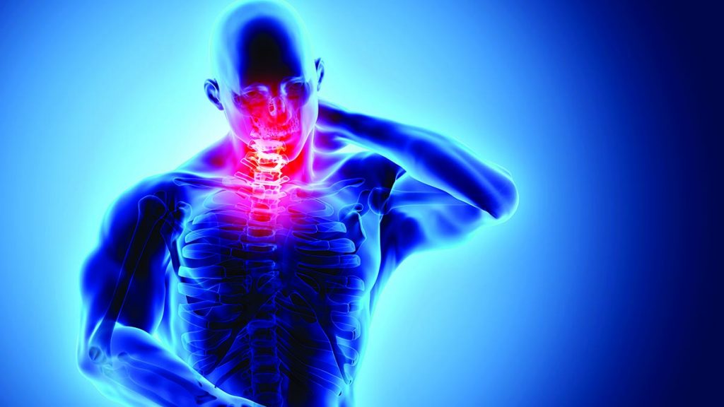
Do you know how to check if you have forward head posture (FHP)?
There are different options for assessing head and neck alignment – from a simple home visual test to a quantitative measurement of the craniovertebral angle (CVA) or a professional postural assessment.
Each method serves a different purpose. Some are useful for simple screening at home, while others are used by healthcare professionals to measure posture, assess neck movement, or investigate symptoms.
A systematic review examined different non-radiographic methods used to measure forward head posture, including photographic and angle-based assessments.
The review found that the reliability of these methods ranged from moderate to excellent, while the evidence for their validity was less conclusive.
In this article, I will describe 8 ways to test and assess forward head posture. Moreover, for each method, I will discuss what it measures and when diagnostic imaging and tests are needed.
Article Contents
- What Is Forward Head Posture?
- How Is Forward Head Posture Tested?
- Visual Side-View Posture Check
- Wall-Based Head Position Check
- Side-View Photograph
- Craniovertebral Angle (CVA) Measurement
- Plumb-Line Assessment
- Cervical Range-of-Motion Assessment
- Physical Therapist Postural Assessment
- Imaging and Neurological Tests When Clinically Indicated
- Which Forward Head Posture Tests Can You Do at Home?
- When Should You See a Healthcare Professional?
- Frequently Asked Questions
- Bottom Line
- References
What Is Forward Head Posture?
Forward head posture refers to a position of the head in which it is more forward in relation to the trunk than one would expect to see in a more neutral posture.
It can be measured by observing anatomical landmarks on the head, neck and upper trunk from the side view. Researchers and healthcare professionals have developed several ways to measure this including calculating the craniovertebral angle.
Keep in mind that your posture is not always the same. Your posture can vary greatly depending on if you are sitting, standing, walking, working at a computer, using a cell phone or engaging in other activities.
This means that a single casual photograph may not tell you the whole story. For a more useful assessment, the photograph should be taken from a standardized position.
If you are also experiencing stiffness, discomfort, headaches or other concerns, our guide to forward head posture symptoms can help you understand the broader signs that may occur with postural problems.
How Is Forward Head Posture Tested?
There is no single measurement for forward head posture.
Rather, there are multiple methods for assessing forward head posture, including:
- visual inspection,
- a wall test,
- standardized photographs,
- craniovertebral angle measurement,
- plumb-line assessment,
- cervical range of motion,
- evaluation by a physical therapist,
- imaging or neurological examination for other associated diagnoses
The most appropriate method depends on what you are trying to determine. A wall test or photograph may be useful for home screening, while CVA measurement and professional assessment provide more objective information.
For instance, the wall test is useful for detecting postural deviation at home, whereas the CVA measurement is more objective.
Finally, evaluation by a physical therapist allows for an analysis incorporating the effects of posture on other components of physical health.
Visual Side-View Posture Check (Test #1)
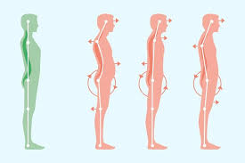
One of the easiest ways to screen forward head posture is to observe your head and neck from the side.
Stand naturally (don’t over-do your posture or try to make your chin stick out). Ask someone to observe your posture from the side and take note of the relationship of your ear, shoulder and upper trunk. You may notice that your head looks like it’s sitting further forward than your trunk.
A quick visual can be helpful, but should not be considered a diagnostic tool.
Studies have shown that simple visual and other basic forms of assessment can be helpful for determining head posture but standardized assessment tools are more reliable.
What can a simple visual tell us?
A visual can help answer these questions:
- Does my head appear noticeably farther forward than my upper trunk?
- Does this change when I’m relaxed?
- Do I look different when I sit versus stand?
- Is there an obvious difference between my natural posture and a neutral/comfortable posture?
If you notice a possible problem, you can follow up with a more objective measurement.
Wall-Based Head Position Check (Test #2)
A wall-based test is another simple screening procedure that you can perform at home.
You have to stand facing a wall, placing your heels, buttocks, and shoulder blades as close to it as possible in a comfortable position
Do not force your body into the position.
Now, look at where the back of your head is in relation to the wall.
If the back of your head does not comfortably touch the wall while the rest of your body is positioned as described, this may indicate that your head is positioned relatively far forward. However, this finding alone does not establish that you have forward head posture.
The study conducted in 2025 assessed the qualitative wall-based test, in which lack of contact between the back of the head and the wall was considered a positive result.
A 2025 study evaluated this wall-based method in healthy young adults and compared it with photographic CVA measurements. The wall test provided useful screening information, but its predictive value was limited compared with the CVA-based criteria used in the study.
Thus, it is better to consider a wall test as a screening procedure rather than a diagnostic one.
If you want a more detailed home assessment, see our guide on how to check your forward head posture at home.
Side-View Photograph (Test #3)
Taking a photograph from the side will probably make it easier to judge your head position than trying to see your posture from the front.
For a better comparison:
- Stand in your normal, relaxed posture.
- Make sure the camera is at about shoulder level.
- Take the photograph from directly to your side.
- Keep the camera far enough away to prevent too much perspective distortion.
- Don’t correct your posture deliberately for the photograph.
- Take more than one photograph if you have to.
Researchers often use photographs to measure head and neck alignment, for example in calculating the craniovertebral angle. Taking photographs can be a reliable method for measuring these angles if the procedure is standardized.
The important word over here is standardized.
Changing your camera angle, posture, or head position between photographs can make the measurements much less reliable and difficult to compare.
A photograph can show you what your posture is like, but it doesn’t in itself tell you that you have a medical condition, or that your posture is the cause of your symptoms.
Craniovertebral Angle (CVA) Measurement (Test #4)
CVA is one of the most commonly studied measurements for quantifying forward head posture in research and clinical assessment.
The craniovertebral angle (CVA) is one of the most common quantitative measurements for forward head posture.
Typically, it is measured from a lateral photograph using anatomical reference points near the seventh cervical vertebra (C7) and the head.
It provides a numeric value by which the position of the head relative to the cervical spine and upper trunk can be described.
Studies have demonstrated that it can reliably be measured using standardized methodologies. For example, one clinical trial found very high intra- and inter-rater reliability when measuring CVA using an electronic head-posture device.
Other studies have examined photographic CVA measurements and have shown that it can be used to differentiate various levels of forward head posture.
However, proposed cut-off values differ across studies, meaning there are no established criteria that might consistently be used as a diagnostic cutoff across populations.
This is an important distinction.
You may find that different websites propose that a certain value of CVA inherently designates “normal” forward head posture or otherwise. Research is more nuanced than that due to variations between equipment and posture, populations, and assessment methodology.
A forward head posture diagnosis should not be based solely on a single home test or photograph. A healthcare professional can consider your posture, symptoms, movement and other findings together when determining whether further assessment is appropriate.
Want to learn how CVA is actually measured?
For a detailed explanation of the landmarks, measurement process and interpretation, see our guide to craniovertebral angle (CVA) measurement for forward head posture evaluation.
Plumb-Line Assessment (Test #5)
A plumb-line assessment provides another way to evaluate body alignment against a vertical reference.
A plumb line is a weighted line that uses gravity to provide a vertical reference. The clinician uses this information to evaluate the relationship between particular body segments and the line.
In the case of forward head posture, the practitioner examines the placement of the head and the cervical region in relation to the rest of the body’s vertical.
Just like the other methods of determining a postural deviation, the results are dependent on the positioning and the protocol used.
For that reason, it is better to view the plumb line as an alignment assessment tool rather than a standalone diagnostic test for forward head posture.
You can learn more about this method in our guide to the plumb-line test for forward head posture.
Cervical Range-of-Motion Assessment (Test #6)
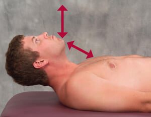
A healthcare professional may also assess the range of motion of your neck in various directions.
This may include:
- flexion
- extension
- rotation
- side bending
The goal is not to determine an “ideal” value for each direction of motion, but rather to use range-of-motion testing as a tool to aid in understanding your neck’s mobility, restriction, and any potential pain or restrictions that may impact your overall assessment of your neck.
Cervical range-of-motion measurements have been shown to be reliable when using standardized procedures.
However, restricted range of motion of the neck alone is not sufficient evidence for forward head posture.
Similarly, a relatively large range of motion is not necessarily an indicator of ideal head and neck posture.
Range-of-motion testing should, therefore, be used as a small part of a comprehensive assessment of your neck.
Physical Therapist Postural Assessment (Test # 7)
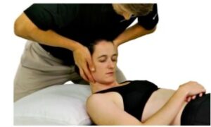
If you want a more thorough assessment, a physical therapist can evaluate your posture and neck mobility rather than relying on a single measurement.
Based on your condition and symptoms, the evaluation may consist of:
- inspection of the head and neck position
- shoulders and posture of the upper back
- range of motion of the cervical spine
- movement patterns
- muscle performance
- pain or restriction evaluation
- functional activities that may be affected by posture
- clinical postural measurements
Research suggests that standardized posture-analysis techniques can provide reliable measurements of forward head posture.
A physical therapist’s evaluation can also help identify if there’s a difference between a postural assessment and another condition that may need further examination and treatment.
For more information, see our guide on how a physical therapist evaluates forward head posture.
Imaging and Neurological Tests When Clinically Indicated (Test #8)
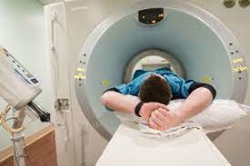
CT scans, MRI, EMG and myelography can sometimes show up in the middle of a list describing “forward head tests.”
This may give the impression that these are routine assessment that most people should get if they have postural FHP.
These are medical investigations used to look for specific structural, neurological, or other clinical problems.
Let me break down the differences now.
X-rays and CT scans
These are imaging techniques good for capturing the state of one’s bones, among other things.
The results of such a diagnostic procedure can help a physician identify any potential irregularity, injury, etc., so it can be addressed accordingly.
MRI
MRI scans are used when there is a need to investigate soft tissues.
MRI can be useful when there is concern about abnormalities involving the discs, nerves, or other soft tissues.
Again, it is a procedure used to address specific concerns that go beyond postural FHP.
EMG
An electromyography is a test used to see how one’s muscles and nerves function.
It may be considered when symptoms such as numbness, tingling, weakness, or other findings raise concern about nerve or muscle dysfunction.
Not exactly a routine procedure for the sake of assessing forward head posture.
Myelography
This is a type of diagnostic imaging usually done with the assistance of a CT scan, employing the use of a special dye in order to further illustrate the spine and nerve roots.
It is once again helpful in the case when there are particular concerns, rather than a general postural assessment.
Now, here is the key point:
If your only concern is that your head appears to sit too far forward, a postural assessment is generally more relevant than immediately pursuing advanced imaging.
However, if there are other concerns, a physician will be able to advise you on what course of action would be best.
Which Forward Head Posture Tests Can You Do at Home?
Several basic assessments can be performed at home, but their purpose is mainly screening and observation, not diagnosis.
| Assessment | Can You Do It at Home? | What It Measures | Diagnostic by Itself? |
|---|---|---|---|
| Visual side-view check | Yes | General head position | No |
| Wall-based check | Yes | Relative head position | No |
| Side-view photograph | Yes | Postural alignment | No |
| CVA measurement | Possible | Quantitative head position | No |
| Plumb-line assessment | Possible | Vertical body alignment | No |
| Cervical ROM assessment | Better performed professionally | Neck movement and limitations | No |
| Physical therapist assessment | No | Posture, movement and function | Professional assessment |
| CT/MRI/EMG | No | Specific structural or neurological findings | Used when clinically indicated |
This distinction is important because testing your posture is not the same thing as diagnosing a medical condition.
What Is the Most Useful Test for Forward Head Posture?
There is not one test that is universally best for every person.
If you simply want to screen your posture at home, a standardized side-view photograph can be a useful starting point.
If you want a numerical measurement, CVA is one of the most widely studied approaches for quantifying forward head posture.
Studies of its reliability are generally positive, although different studies have used different criteria for defining the angle and different cutoffs.
If you have pain, decreased range of motion, weakness, numbness, or other symptoms, an in-person exam may be much more informative than either of the above.
When Should You See a Healthcare Professional?
Consider having a healthcare evaluation if your neck or posture concerns are associated or complicated by other symptoms that persist, worsen or interfere with activities.
This includes:
- persistent or significant neck pain
- recurrent headaches associated with your neck symptoms
- numbness or tingling
- unexplained muscle weakness
- marked restriction of neck movement
- symptoms that began after an injury
- poor balance or coordination
- pain that repeatedly interferes with sleep
A healthcare professional can determine if your symptoms are related to your posture or if other conditions need to be evaluated.
A home posture test should not be used to rule out other medical problems.
Frequently Asked Questions:
Can I test for forward head posture at home?
Yes—you can make a rough assessment of your posture by viewing it from the side, performing a wall check, or taking a photograph. These methods can help you identify a possible postural deviation, but they should not be treated as a diagnosis.
How can I check if I have forward head posture?
The first step is to view your posture from a side view and assess your head position. You can also do a wall check, or take a photo.
To quantify it, a measurement of your craniovertebral angle can be taken from a standardised photograph.
What is the most common test for forward head posture?
There is no single test that is universally used for everyone. Visual assessment, standardized photographs, and craniovertebral angle measurement are among the methods studied for assessing FHP.
A systematic review of 21 studies found that the reliability of non-radiographic measurement methods ranged from moderate to excellent, although the evidence for validity was less conclusive.
What is a craniovertebral angle?
The craniovertebral angle (CVA) is an angle measured from a side-view photograph using landmarks around the ear and the seventh cervical vertebra (C7). It provides a quantitative way to describe the forward position of the head relative to the neck and upper trunk.
What CVA angle indicates forward head posture?
There is no magic number for a CVA that indicates forward head posture. Various papers have looked at using a CVA cutoff for differentiating between slight, moderate and severe forward head posture, with varying results.
One study by Mostafaee et al. identified a CVA cutoff of 45.5° for distinguishing slight from moderate-to-severe FHP within its study population.
As such, I would be cautious of using a CVA cutoff to determine forward head posture. It should be considered as part of a wider assessment of your posture.
Can a wall test detect forward head posture?
Yes, a wall test can give an indication of forward head posture. However, research by Mostafaee et al. indicated that it was not as reliable or accurate as some other methods of quantifying postural deviation, such as measuring the CVA on a photograph.
Can a physical therapist diagnose forward head posture?
A physical therapist can assess your posture as part of a broader physical examination. This might include a visual assessment, range of motion tests, motion tests, and objective measurements such as the CVA.
Do I need an X-ray or MRI to diagnose forward head posture?
Not necessarily. Getting an X-ray, CT or MRI scan is unlikely to be indicated simply to check for forward head posture. The purpose of these tests is to look for other musculoskeletal issues, which may be recommended when your symptoms or physical examination suggest that further investigation is needed.
Can forward head posture be diagnosed from a photograph?
A standardized photograph can be used to assess and measure aspects of forward head posture, particularly when calculating the craniovertebral angle. However, a photograph alone cannot establish the cause of the posture or diagnose every possible neck condition.

Bottom Line
If you want to know whether you have forward head posture, you’ll probably not need an MRI, CT scan or some kind of advanced medical test.
A basic glance at a lateral (side) view can be helpful, a wall test, standardized photograph, or quantitative measurement can provide additional information. If you want a number, the craniovertebral angle (CVA) is a fairly well-established method of calculating it.
If you have significant pain, marked loss of range of motion, neurological symptoms, or other concerning findings, a physical therapist or other medical professional would be able to perform a more comprehensive assessment.
The important point is that no single home test or measurement tells the entire story. Use these assessments as tools for understanding your posture, and seek professional evaluation when symptoms or other concerns warrant it.
Medical Disclaimer:
This article is for informational purposes only. Home posture checks and measurements are not a substitute for an assessment by a qualified healthcare professional and cannot diagnose an underlying medical condition.
If you have persistent or severe pain, numbness, weakness, loss of balance or coordination, symptoms following an injury, or other concerning symptoms, consult an appropriate healthcare professional.
References:
- Reliability and Validity of Non-radiographic Methods of Forward Head Posture Measurement: A Systematic Review — PubMed
- Clinical Measurement of Craniovertebral Angle by Electronic Head Posture Instrument: A Test of Reliability and Validity — PubMed
- Examining Accuracy of and Determining the Best Cutoff Point for Photographic-Based Postural Angles to Discriminate Between Slight and Moderate-to-Severe Forward Head Posture — PubMed
- Validity of a Qualitative Visual Method for Diagnosing Forward Head Posture — PubMed
- Reliability of Forward Head Posture Evaluation While Sitting, Standing, Walking and Running — PubMed
- Reliability of Measuring Forward Head Posture in a Clinical Setting — PubMed
- Measurement of Craniovertebral Angle by the Modified Head Posture Spinal Curvature Instrument: A Reliability and Validity Study — PubMed
- Why Trust Us? https://bestforwardheadposturefix.com/why-trust-us/